

Walk into any modern hospital, and you’ll find a medical imaging department. Medical imaging uses x-rays, magnetic resonance imaging (MRI), and other arcane-sounding methods like positron emission tomography (PET) to image the body’s interior for analysis and diagnosis. To a non-specialist, these techniques can sound almost otherwordly. But in one way or another, these technologies rely on natural phenomena, including radiation, to do their thing.
Now a new study suggests that the Universe’s naturally occurring radiation could be used in medical imaging and could be particularly useful when it comes to COVID-19. The type of radiation in question is cosmic rays.
The term “cosmic rays” is one of science’s historical misnomers. Cosmic rays are not actually rays, but rather high-energy particles, usually protons. They can originate from the Sun, from somewhere else in the Milky Way, or from even further beyond, from some distant location in the Universe.
When these high-energy particles reach us, they collide and interact with Earth’s atmosphere, producing muons. A muon is similar to an electron but with a much greater mass. Muons don’t last long and decay after a couple of microseconds. But they travel at relativistic speeds and cover a lot of distance before they decay. They’re so energetic that many of them reach Earth’s surface. According to the paper’s authors, the muons that reach the Earth could be used in a medical imaging technique called radiography.
The researchers say that muons could be used “…to monitor bulk aspects of human anatomy.” Since the shower of muons is continuous, the technique could be used to monitor changes over time, down to an hour’s length if necessary. The authors point out how useful this could be in our current times. “This could provide hourly readouts of parameters such as lung density with sufficient sensitivity to detect time changes in inflammation of the lungs in e.g. Covid patients,” they write.
One of the effects of COVID-19 concerns lung tissue density. As the immune system tries to fight the infection, the density of the infected lung tissue increases. This increase is associated with severity, and the greater the increase in lung tissue density, the greater the likelihood of the patient requiring intensive care or even respirators. Being able to monitor the change in density could be a huge boost in the treatment of COVID-19.
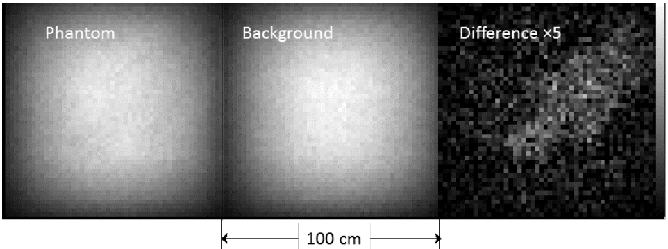
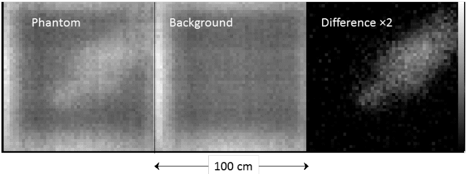
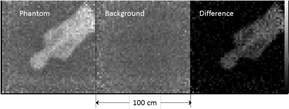
The researchers used what’s called a “human phantom” in medical imaging. A human phantom is something that stands in for a human body in research. It helps researchers determine if their methods are working correctly. In this work, they installed muon trackers both above and below the phantom. The system measures the trajectories of the incoming and the outgoing muons after they pass through the phantom. Then they measure the angular distribution of the outgoing muons, which allows them to measure the density of the tissue they passed through.
The detectors above and below the phantom allowed the team to work with three different types of radiography to measure the muon tracks. They are transmission, stopping, and multiple scattering.
Transmission radiography measures the muons as they are transmitted through an object.
Stopping radiography measures and projects the tracks of the muons as they enter the phantom but not as they leave.
Multiple scattering measures the difference in angle between incoming and outgoing muons.
Each of the above images used an exposure time of 24 hours, and the detectors they used are not the most sensitive. But this is preliminary work, and the team says that much more sensitive detectors could be used, which would shorten the required exposure time and raise the image quality. That’s what creates the potential for near-continuous monitoring of a patient using only background muon radiation.
“Lower mass higher efficiency detectors, such as wire chambers, could provide higher quality images in considerably shorter exposure times with much better signal to noise. Changes in non-ambulatory patients may be able to be
continuously monitored using only background radiation,” they write in their conclusion.
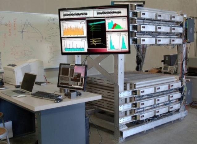
The title of the paper outlining this potential is “Cosmic Ray Radiography of a Human Phantom.” The authors are from the Los Alamos National Laboratory and used that facility’s Mini Muon Tracker to examine the human phantom. The paper is available on the pre-press site arxiv.org and hasn’t yet been peer-reviewed.